A summary of our work on prostate imaging, mostly between 2010 and 2013, around the PhD theses of Soumya Ghose (segmentation) and Jhimli Mitra (multimodal registration), with later work on web tools for prostate MRI. Each topic links to the related papers.
Prostate segmentation
Delineating the prostate is needed for biopsy guidance, treatment planning and volume estimation, but its boundary is often weak, especially in transrectal ultrasound (TRUS). We first surveyed segmentation methods across ultrasound, MRI and CT.
Shape and appearance models
Our early work extended Active Appearance Models with texture: AAM propagation guided by texture, local binary patterns, and quadrature phase information to build statistical shape and appearance models that are robust to the low contrast of TRUS.
Learning shape and probability priors
We then combined statistical shape models with probability priors learned by supervised classifiers, using multiple mean models and spectral clustering to capture shape variability, and embedded them in a supervised learning framework for automatic segmentation of ultrasound images.
3D segmentation in MRI
For MRI we coupled probabilistic atlases with statistical shape and appearance models, used graph-cut energy minimisation, multiple active appearance models with global registration, and a Mumford–Shah variational model with contour, shape and probability priors. A random forest approach took part in the PROMISE12 challenge.
Multimodal registration for biopsy
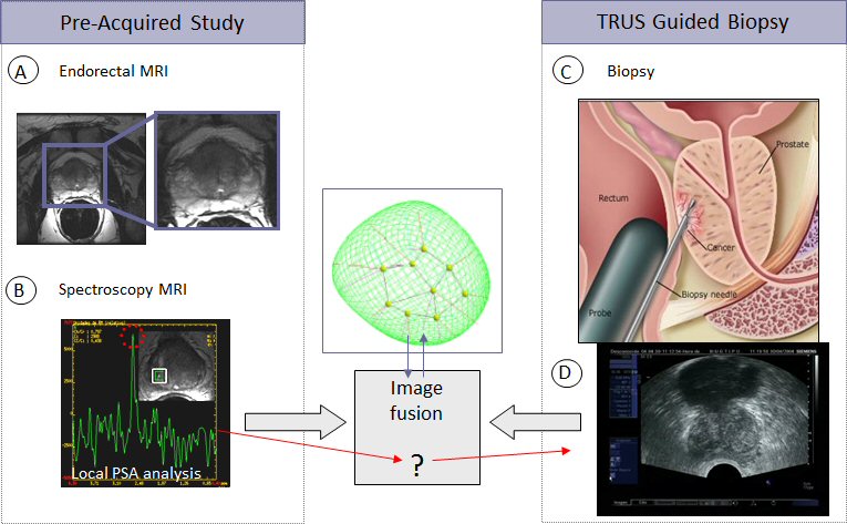
TRUS-guided biopsy is the standard to diagnose prostate cancer, but ultrasound shows tumours poorly, while MRI shows them well. Fusing pre-acquired MRI with the TRUS images during the procedure brings the MRI information to the biopsy needle.
Deformable registration
We compared thin-plate splines with automatic correspondences and b-splines on uniform grids, enhanced b-spline registration with quadrature filters and local energy, and proposed non-linear diffeomorphic and spline-based diffeomorphic frameworks, together with spectral clustering of deformations for fast registration.
Slice correspondence
Before registering, the MR slice that corresponds to the live 2D TRUS slice has to be found. We addressed this with shape-based statistics, joint probability of shape and image similarities, and weighted likelihoods of multiple statistical parameters.
Web tools for prostate MRI
With clinicians, we built ProstateAnalyzer, a web-based application to manage and annotate multiparametric MR images of prostate cancer, studied how XML supports medical imaging web applications, and evaluated the benefits of collaborative work between experts when assessing prostate cancer from MRI.