A summary of my work on brain imaging since 2010, with Xavier Lladó and the ViCOROB neuroimaging team, together with clinical partners. It started with multiple sclerosis and has grown into stroke, brain tumours and brain ageing, moving from classical image analysis to deep learning. Each topic links to the related papers.
Multiple sclerosis
Multiple sclerosis has been the backbone of our neuroimaging work since 2010, together with clinical partners. Over these years the methods have moved from intensity-based and supervised classifiers to deep learning, but the clinical questions have stayed the same.
Lesion segmentation
After reviewing automated approaches, we proposed supervised (BOOST) and unsupervised (FLAIR thresholding) methods and released them as a lesion segmentation toolbox and an SPM12 extension. We later moved to cascaded 3D convolutional networks, one-shot domain adaptation to new scanners with very few labelled images, synthesis of lesions in MRI, and longitudinal recurrent models.
New lesions in follow-up scans
Detecting new or enlarging T2 lesions between visits is key to monitoring treatment. Our pipelines evolved from image subtraction and deformation-field features to fully convolutional networks, and we evaluated decision-support systems for active lesions and the use of synthetic T1-w images.
Lesions and brain volumetry
White-matter lesions bias tissue and structure segmentation, and therefore atrophy measurements. We quantified this effect across several tools, proposed lesion filling, and designed tissue and sub-cortical segmentation methods that are robust to lesions.
Clinical prediction
Recent work uses deep learning on clinical MRI to stratify patients, to explain disability through attention maps, and to predict progression independent of relapse activity from the first demyelinating event.
Brain tissue, structures and atrophy
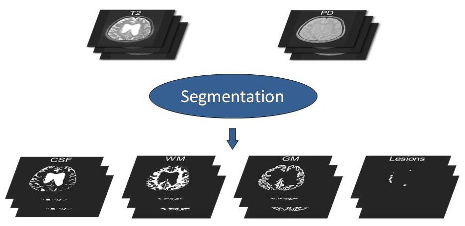
Accurate brain measurements underpin many neurological studies. We reviewed atlas-based segmentation, brain structure segmentation and deep learning for brain MRI, and compared ten tissue segmentation methods on revisited IBSR annotations.
Our methods cover brain extraction (MARGA and PARIETAL), tissue segmentation with patch-based networks, and sub-cortical structure segmentation combining spatial and deep features, with supervised and transductive domain adaptation so that models work on new data with little user interaction.
For longitudinal atrophy we studied registration and intensity standardisation, built datasets for evaluating atrophy measurements, and improved atrophy quantification by training on automated labels with tissue-similarity priors.
Infant and fetal brain
We have also segmented the developing brain: six-month infant tissues, the infant cerebellum, fetal tissues, and the Sylvian fissure in ultrasound of preterm infants.
Stroke and cerebrovascular disease
Stroke is now one of our main lines, developed together with clinical stroke teams.
Ischaemic lesions
We segment the acute ischaemic core in CT perfusion and acute and sub-acute lesions in multimodal MRI, and took part in the ISLES challenges, contributing to the DeepISLES model.
Large vessel occlusion
Detecting a large vessel occlusion quickly in CT angiography decides who goes to thrombectomy. We have explored how to combine 3D vasculature with CTA, how to encode 3D information in 2D feature maps, window self-attention with volumetric refinement, and localisation guided by the Circle of Willis to detect and classify occlusions.
Circle of Willis
Through the TopCoW challenges we worked on topology-aware, multiclass segmentation of the Circle of Willis in MRA and CTA.
Intracerebral haemorrhage
We segment haemorrhagic lesions with 3D U-Nets and semi-supervised learning, and predict hematoma expansion from the admission CT with end-to-end deep learning, including the use of synthesised follow-up images.
Brain tumours
We have taken part in the BraTS challenges since 2018: tumour segmentation and survival prediction, uncertainty quantification (QU-BraTS), segmentation for a sub-Saharan African population, meningioma segmentation for radiotherapy planning, and synthesis of missing MRI modalities.
Brain age and other applications
Our most recent line models brain ageing, with age-conditioned brain atlases for brain age prediction. Other applications include lupus white-matter lesions, white-matter hyperintensities, autism spectrum disorder from structural and functional MRI, and traumatic brain injury. We also study how ensembles reveal confounding in medical imaging models.